
Publications
Papers
Sakadžić, S., Mandeville, E.T., Gagnon, L., Musacchia, J.J., Yaseen, M.A., Yücel, M.A., Lefebvre, J., Lesage, F., Dale, A.M., Ekermann-Haerter, K., Ayata, C., Srinivasan, V.J., Lo, E.H., Devor, A., Boas, D.A., "Large arteriolar component of oxygen delivery implies safe margin of oxygen supply to cerebral tissue" Nature Communications 5, 6734 (2014).
ABSTRACT:
What is the organization of cerebral microvascular oxygenation and morphology that allows adequate tissue oxygenation at different activity levels? We address this question in the mouse cerebral cortex using microscopic imaging of intravascular O2 partial pressure and blood flow combined with numerical modelling. Here we show that parenchymal arterioles are responsible for 50% of the extracted O2 at baseline activity, and the majority of the remaining O2 exchange takes place within the first few capillary branches. Most capillaries release little O2 at baseline acting as an O2 reserve that is recruited during increased neuronal activity or decreased blood flow. Our results challenge the common perception that capil- laries are the major site of O2 delivery to cerebral tissue. The understanding of oxygenation distribution along arterio-capillary paths may have profound implications for the interpreta- tion of blood-oxygen-level dependent (BOLD) contrast in functional magnetic resonance imaging and for evaluating microvascular O2 delivery capacity to support cerebral tissue in disease.
CO-AUTHOR CONTRIBUTION:
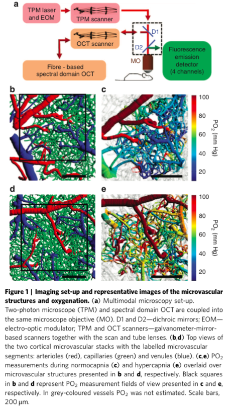
- Performed vessel segmentation to prepare image stacks for analysis presented in subsequent figures (most literally shown in panels b-e in Figure 1)
- Modified MATLAB code used to perform cleaning (filtering, masking, etc)
Gagnon, L., Sakadžić, S., Lesage, F., Musacchia, J.J., Lefebvre, J., Fang, Q., Yücel, M.A., Evans, K.C., Mandeville, E.T., Cohen-Adad, J., Polimeni, J.R., Yaseen, M.A., Lo, E.H., Greve, D.N., Buxton, R.B., Dale, A.M., Devor, A., Boas, D.A. “Quantifying the microvascular origin of BOLD-fMRI from first principles with two-photon microscopy and an oxygen-sensitive nanoprobe” The Journal of Neuroscience - Accepted, (January, 2015).